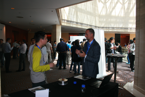
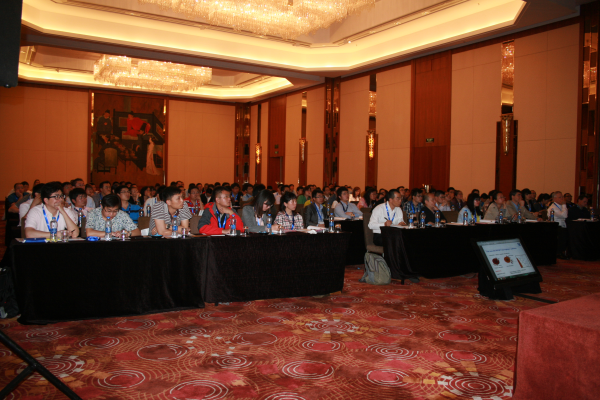
A few days ago I was invited by Merck Millipore to contribute to the Forum they organized in Shanghai. Besides being one of the speakers –more details below-, SEPMAG has also been involved as sponsor. We believed the direct contact with IVD-magnetic beads users in China, as well as with the technical worldwide contributors, is the only way to still push forward this technology.
After few days to catch-up with the day-to-day, let’s me share some of my notes about the presentations. I have tried to organize the following paragraphs by subjects, not just by the order of the programs.
Let us review first some of the academic authors who introduced us new trends on immunomagnetic assays. Dr. John X.J. Zhang from Shanghai Jiao Tong University, explained how magnetic cell separation was particularly valuable for rare cell separation such as for Circulating Tumor Cells (CTC). He reviewed the rare cancer cell sorting technologies based on magnetic-activated and immunomagnetic assay over multiple scales. He discussed macroscopic cell detection system, followed by the scaling laws on miniaturization towards microfluidic chips. He introduced recent work that integrates micro-magnets in separation experiment, that demonstrated >90% capture rates for four different cell lines (COLO 205, PC3, SK-BR-3, MCF-7) with 15% increase in detection sensitivity and 10% increase in detection stability compared with glass slide without patterned array.
A second academic contributor was Dr. Maxime Nikitin from the Russian Academy of Sciences. He described two different developments: a rapid quantitative detection of biomolecules by MP-based immunochromatography and a non-invasive detection of magnetic nanoparticle in blood and organs of small experimental animals. In the first magnetic particles used as nanolabels of biochemical reactions are trapped inside a 3D fiber. The sample under test is exposed to an AC magnetic field at two frequencies, leading to a robust reading, with high signal-to-noise ratio and a remarkable wide linear range. The report detection limit was as low as 25 pg/mL for a prostate-specific antigen. A second method he explained was a non-invasive detection of magnetic nano-particles in blood and organs of small experimental animals by non-linear magnetization. Finally he also mention the advantages of using proteinaceous ‘molecular glues based on the barnase-barstar system.
Coupling of biomarkers was other of the subjects widely discussed, both at the talks and at the numerous coffee breaks. Dr. Steve Henry, from KODE Biotech introduces KODE technology, an amphipathic molecule that self assembles as coating onto surfaces. He describes as it can enable users to rapidly modify biological and non-biological surfaces with virtually any biological or non-biological material. According Dr. Henry this construct not only can reduce costs and accelerate R&D, but it can also facilitate and expand research and development opportunities, particularity when used in conjunction with microspheres. A second contributor, Dr. Richard Vidal, Director of Merck Millipore, described new microspheres development for one-step antibody coupling are hydrophobic magnetic microspheres for passive adsorption method, low COOH loading microspheres for the facilitated (forced) adsorption method, the Tosyl, Chloromethyl and epoxy modified for covalent coupling.
More specific in biomarkers for immunomagnetic assays Dr. Michael Mansfield, from EMD Millipore describes with detail how antibodies used for immunohistochemistry of the tau protein –a biomarker for Alzheimer’s disease- were screened to identify the best suited for ELISA. In first round were characterized for their ability to recognize tau protein in a direct ELISA assay. They were then screened in sandwich assay using different capture antibodies on the ELISA plate and different sources of tau protein. As anticipated, the antibodies exhibited different levels of reactivity. A set was chosen that was best suited to detecting tau protein in a sandwich assay. While may failed, certain worked adequately in that they produced good signals without generating high background. These antibodies were used for the development of an assay based on magnetic microspheres. The immunoreagents resulting from this series of screening steps can now be developed into an assay format that will allow for the rapid measurement of the tau protein in cerebrospinal fluid.
As SEPMAG’s Chief Scientific Officer I told about how to avoid 5 critical mistakes when using Biomagnetic Separation in CLIA IVD-kits Manufacture. After 10 years helping leading IVD companies to implement Biomagnetic Separation Processes, we have identified five critical mistakes that delay projects, cause economic losses and, sometimes, endanger production. We will discuss with more detail in further Posts, but the list we have elaborated comprises (1) Blaming the magnetic beads for a deficient separation process, (2) Using bigger magnets to avoid losses (3) Defining the process by separation time alone (4) Neglecting process scalability, and (5) Inappropriate safety precautions when working with magnetic fields..
In a next post I’ll review the contributions about lateral flow and immunoturbidimetry, as well as the discussion about readers and microfluidics.